LPi1-2 [FBbt_00052850]

LPi1-2
ID: FBbt_00052850
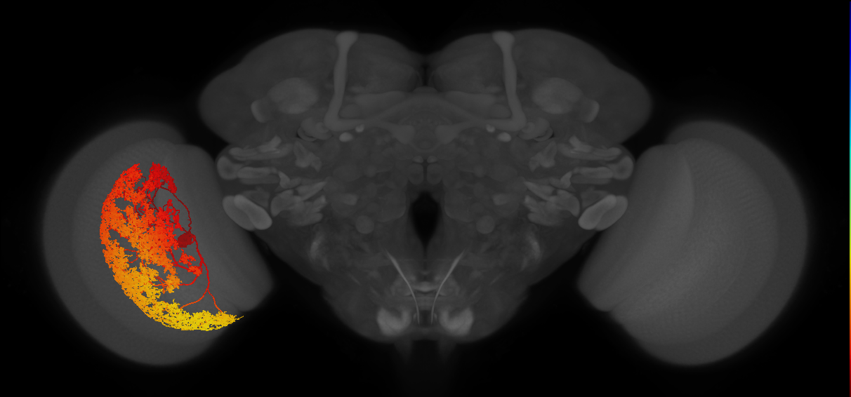
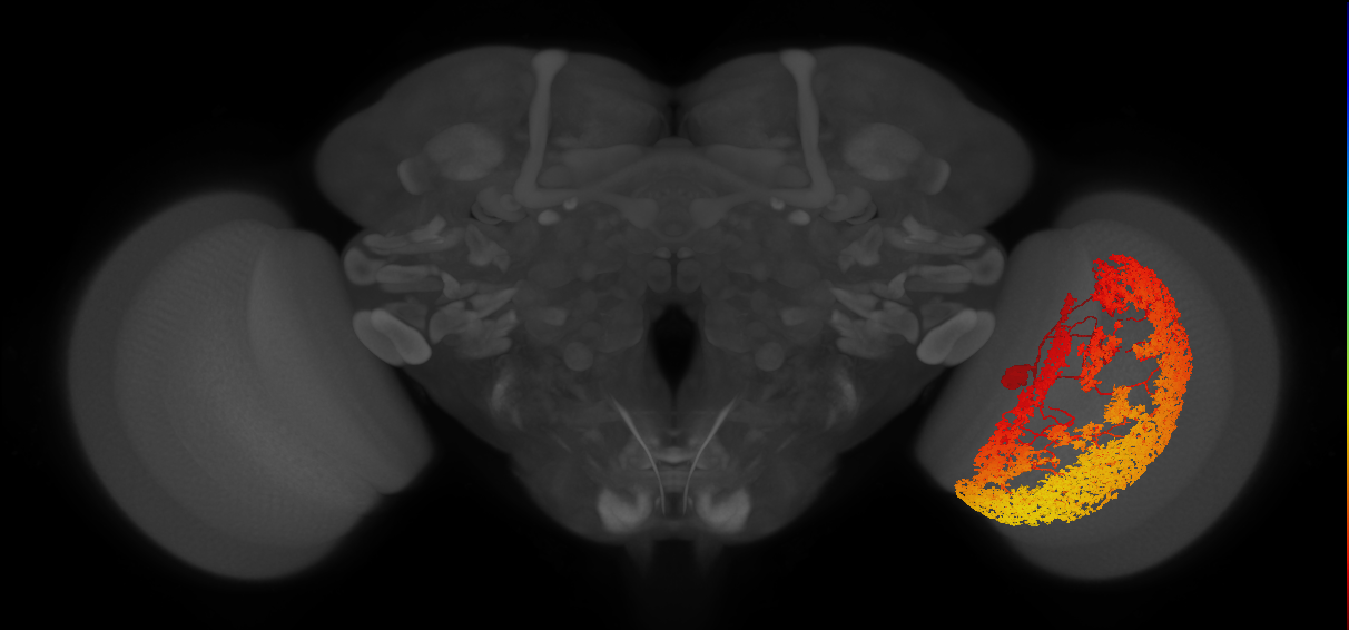
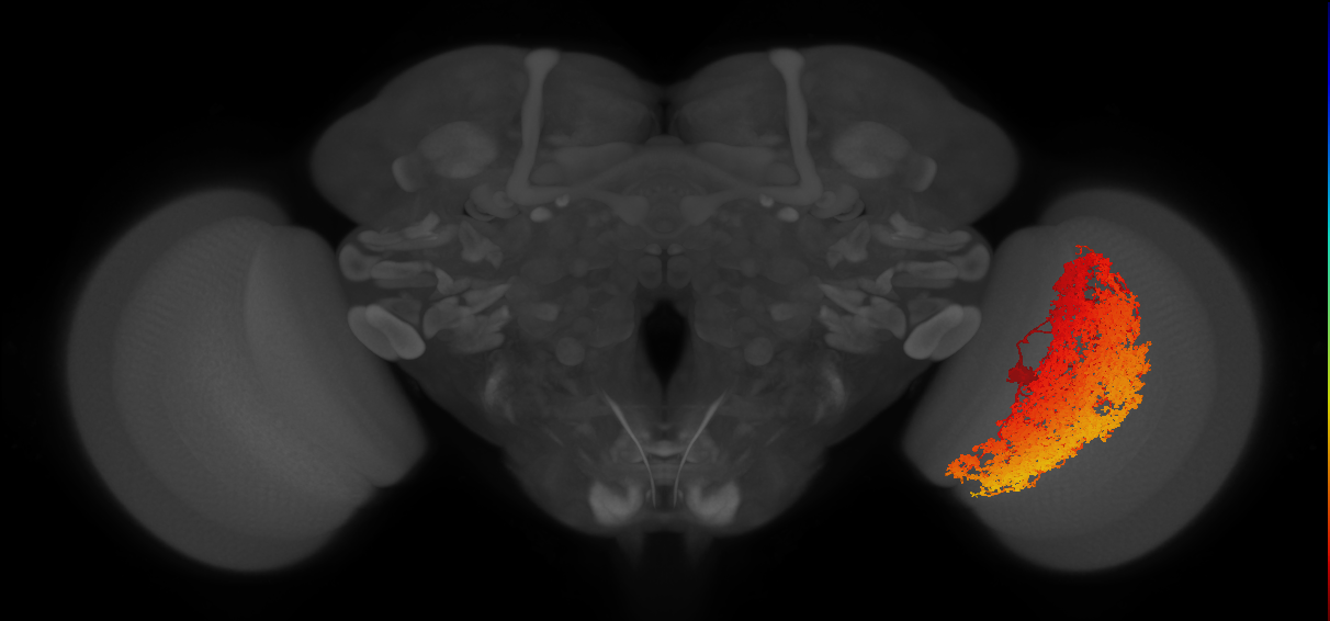
Intrinsic neuron of the lobula plate that has its synaptic terminals throughout, but restricted to lobula plate layers 1 and 2, receiving input in layer 1 and sending output to layer 2 (Shinomiya et al., 2022. The majority of its input is from T4a and T5a cells (Shinomiya et al., 2022. It has its soma in the lobula plate cell body rind (Shinomiya et al., 2022. There are two of these cells per optic lobe and they tile the lobula plate in a jigsaw-like pattern (Matsliah et al., 2024; Dorkenwald et al., 2024; Schlegel et al., 2024. (Dorkenwald et al., 2024, Shinomiya et al., 2022, Matsliah et al., 2024)
Matsliah et al., 2024 - FBrf0260545 nomenclature scheme unrelated to layer innervation.
Open in VFB 3D Browser →Classification
Relationships
- has soma location: cell body rind of adult lobula plate
- receives synaptic input from neuron: T4a neuron, T5a neuron
- receives synaptic input throughout: lobula plate layer 1
- sends synaptic output throughout: lobula plate layer 2
Alternative Names
| Synonym | Scope | Reference |
|---|---|---|
| LPi14 | related synonym | Bates et al., 2025, Schlegel et al., 2024 |
| LPi12 | related synonym | Berg et al., 2025, name_in_male-cns, Nern et al., 2025 |
| LPi14 | exact synonym | Matsliah et al., 2024 |
| LPi1-2 | exact synonym | Shinomiya et al., 2022 |
Downloads
Image files aligned to LPi12_L (MaleCNS:13310):
Image files aligned to LPi12_R (MaleCNS:10436):
Image files aligned to LPi12_R (MaleCNS:10308):
Image files aligned to LPi1-2:
Image files aligned to LPi1-2:
Image files aligned to LPi1-2:
Image files aligned to LPi1-2:
Image files aligned to LPi12_R (JRC_OpticLobe:10436):
Image files aligned to LPi12_R (MaleCNS:10436):
Image files aligned to LPi12_R (MaleCNS:10308):
Graphs For
References
Feedback
Was this page helpful?
Glad to hear it! Please tell us how we can improve.
Sorry to hear that. Please tell us how we can improve.